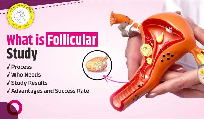
When trying to conceive, understanding the intricacies of your reproductive health can make a significant difference. One such vital tool in fertility treatments is a follicular study—a series of ultrasound scans that monitor the growth and development of ovarian follicles.
This simple yet effective method helps determine the best time for conception, whether naturally or through assisted reproductive techniques. There are many of information that is provided by the Follicular Study Report like the Antral Follicle Count, Follicular Size, Dominant Follicle, Endometrial Pattern/Thickness and so on.
Let’s have a detailed discussion about the Follicular Study!
What is Follicular Study?
In this Article
Follicle monitoring, sometimes called a follicular study, is crucial for evaluating and treating infertility. A sequence of ultrasound scans monitors the growth of ovarian follicles—tiny sacs in the ovaries where eggs mature. Understanding a woman’s ovulation cycle depends on this study. It is frequently utilised in assisted reproductive technologies like IUI (Intrauterine insemination) and (in-vitro fertilization) IVF, as well as in natural conception planning.
Follicular Study Scan: How It Works
A sequence of vaginal ultrasound scans known as “follicle tracking” takes five to ten minutes to complete. Beginning on the ninth day of the cycle, the follicles begin to grow. Until the follicles have vanished and ovulation has begun, the scans continue. It is then recommended that couples engage in sexual activity. By optimising the time of the sperm and egg’s meeting, it aids in conception.
The doctor examines the follicles’ size. Additionally, they measure the endometrium’s (the uterine wall) thickness. A Doppler scan may also be used by the physician to examine the endometrium and follicular blood flow. A healthy endometrium is thought to be necessary for a successful pregnancy, and a mature follicle typically measures 18 to 25 mm.
Importance of Follicular Study in Fertility Treatment
| Aspect | Details |
| Tracking Ovulation | Identifies the timing of ovulation for optimizing natural conception, IUI, or IVF. |
| Assessing Ovarian Response | Monitors ovarian response to fertility medications, ensuring effective and safe stimulation. |
| Preventing Complications | Detects issues like ovarian hyperstimulation syndrome (OHSS) and minimizes risks of multiple pregnancies. |
| Tailoring Treatment | Provides data to customize fertility treatment protocols based on the patient’s response. |
| Improving Success Rates | Ensures procedures like IUI or egg retrieval are timed precisely, increasing the likelihood of successful outcomes. |
| Diagnosing Anomalies | Helps identify conditions like anovulation (lack of ovulation) or poor follicular growth, guiding further treatment. |
Types of Follicular Studies
| Type of Follicular Study | Description | Advantages | Limitations |
| Transvaginal Scan (TVS) | An internal ultrasound that uses a probe inserted into the vaginal canal to get detailed images of the ovaries. | Most accurate method for monitoring follicular growth; provides clear, detailed images. | May not be suitable for patients uncomfortable with internal procedures. |
| Abdominal Ultrasound | An external ultrasound performed by scanning over the lower abdomen to view the ovaries and uterus. | Non-invasive and more comfortable for some patients. | Less detailed compared to TVS; may not clearly show smaller structures like follicles. |
How is a Follicular Study Performed?
Using transvaginal ultrasonography, a follicular study is carried out by inserting an ultrasound probe into the vagina to provide a clear picture of the uterus and ovaries. Here’s what to anticipate:
1. Baseline Scan
Usually conducted on the second or third day of the menstrual cycle, a baseline ultrasound scan serves as the initial step in the process. The uterine lining and any cysts or follicles at the beginning of the cycle are described in this scan.
2. Monitoring Phase
Depending on the patient’s cycle and the doctor’s procedure, follow-up scans are carried out every one to three days. The sonographer measures the size of the growing follicles and evaluates the thickness and pattern of the uterine lining at each visit.
3. Identifying Ovulation
The purpose is to monitor the development of the dominant follicle(s). A follicle is considered developed and ready for ovulation when it measures between 18 and 22 mm in diameter. If blood tests are performed, the doctor may look for symptoms of approaching ovulation, such as the LH surge (Luteinizing Hormone).
4. Post-Ovulation Scan
Sometimes a scan is performed after the planned ovulation date to establish that the follicle ruptured and ovulation happened.
Key Insights from a Follicular Study Report
A follicular study report provides crucial information about ovarian follicular development, ovulation timing, and overall fertility health. Here are the key components and insights:
| Aspect | Details |
| Follicle Growth | Measures the size and number of follicles during the cycle. Dominant follicles should reach 18-20 mm for ovulation. |
| Ovulation Status | Confirms whether ovulation occurred by detecting follicle rupture or corpus luteum formation. |
| Endometrial Thickness | Evaluates the uterine lining to ensure it is optimal for implantation, typically 7-14 mm during ovulation. |
| Ovarian Response | Assesses how the ovaries respond to natural cycles or stimulation with fertility medications. |
| Cycle Monitoring | Tracks the timeline of follicular growth, helping to determine the most fertile window. |
| Abnormalities | Detects any issues such as anovulation (no ovulation), poor follicular growth, or cyst formation. |
| Timing for Interventions | Guides fertility treatments like IUI or IVF by pinpointing the best time for egg retrieval or insemination. |
Benefits of Follicular Study for Fertility
There are numerous advantages to conducting a follicular study. Some of these include:
1️⃣ Detects Growth Issues :The opportunity to identify follicles that are unable to grow normally.
2️⃣ Tracks Ovulation Problems : It aids the doctor in diagnosing when a follicle matures properly but fails to release an egg.
3️⃣ Identifies Uterine Issues : It gives a platform for identifying uterine issues, which are among the most common causes of conception failure.
4️⃣ Early Diagnosis & Treatment :Other problems could be found and treated earlier, preventing further difficulties.
Understanding a Normal Follicular Study Report
| Aspect | Details | Normal Range/Findings |
| Day of Cycle | The report typically begins on Day 9 or 10 and continues until ovulation. | Follicular growth is monitored from Day 9-14 or until ovulation. |
| Follicle Size | The dominant follicle’s size is measured to determine ovulation readiness. | Dominant follicle reaches 18-20 mm for ovulation. |
| Endometrial Thickness | The thickness of the uterine lining is measured to assess readiness for implantation. | Endometrial thickness should be 7-14 mm during ovulation for implantation. |
| Follicular Growth | Tracks the development of follicles and their progression towards maturity. | Follicles grow 1-3 mm per day leading to ovulation. |
| Ovulation Confirmation | The report confirms ovulation by observing follicular rupture or the formation of a corpus luteum. | Ovulation is confirmed by follicle rupture or fluid collection in the pelvic area. |
| Abnormal Findings | Any abnormalities detected during the study, such as poor follicular growth or cyst formation. | No cysts or abnormal growth, and a single dominant follicle should be observed. |
| Cycle Monitoring | Tracks the timing of ovulation to optimize fertility treatments or conception attempts. | Ovulation typically occurs 24-36 hours after the dominant follicle reaches 18-20 mm. |
Cost of Follicular Study
| Factor | Cost Range (INR) | Details |
| Single Scan (Abdominal Ultrasound) | ₹1,500 – ₹3,000 | Basic abdominal ultrasound to monitor follicles, usually less detailed. |
| Single Scan (Transvaginal Ultrasound) | ₹2,000 – ₹4,500 | More accurate method for monitoring follicular growth. |
| Complete Follicular Study (Multiple Scans) | ₹3,000 – ₹8,000 | Includes multiple scans during the cycle to monitor follicular growth, ovulation, and endometrial development. |
| Medicated Cycle Monitoring | ₹4,000 – ₹10,000 | Includes monitoring of follicles during medicated cycles, with scans and possible hormone tests. |
| Additional Costs | ₹500 – ₹2,000 | Additional charges may apply for hormone tests (like LH, FSH, estradiol), doctor consultations, and medications. |
Conclusion
A follicular study provides insight into the complex interplay of hormones and events that contribute to ovulation. It is essential in fertility examinations and therapies, providing important information about a woman’s reproductive health. So, this was all you needed to know about Follicular Study, we hope this blog helps you understand the follicular study report better. This article explains the importance of follicular scans in IVF, with insights from Dr. Rashmi Prasad, the top IVF Specialist Doctor in Patna.
FAQs
What exactly is a follicular study?
A follicular study is a diagnostic procedure where ultrasound is used to track the growth and development of ovarian follicles. It’s often done to check when ovulation occurs, which helps in timing conception or fertility treatments.
Why do doctors prefer transvaginal ultrasounds for follicular studies?
Transvaginal ultrasounds are preferred because they offer clearer and more detailed images of the ovaries, making it easier for doctors to track the size of the follicles and monitor their progress.
When do I start the follicular study during my menstrual cycle?
Usually, the study starts around Day 9 or 10 of your cycle. You’ll have several scans until ovulation occurs, usually between Day 14 and Day 16, to check for follicle growth and other signs of ovulation.
How many scans do I need during a follicular study?
Typically, you’ll need 3 to 5 scans throughout your cycle. The exact number depends on how your follicles are progressing and whether ovulation has occurred.
What size should my follicles be to ovulate?
For ovulation to happen, the dominant follicle needs to reach a size of 18-20 mm. If it’s smaller, it might need more time to grow, and if it’s too large, it could indicate issues like ovarian cysts.
How can I know for sure that I’ve ovulated?
Ovulation is confirmed when the dominant follicle ruptures and releases an egg. Your doctor might also observe the formation of a corpus luteum or notice fluid in the pelvic area, which are signs that ovulation has occurred.
Is a follicular study only for people undergoing fertility treatment?
Not at all! While follicular studies are commonly used in fertility treatments like IUI or IVF, they are also helpful for women trying to conceive naturally or those with irregular cycles, as they can diagnose ovulation issues or cycle irregularities.
How much does a follicular study cost in India?
The cost can range from ₹1,500 to ₹8,000, depending on the type of ultrasound, the number of scans required, and any additional tests or consultations needed.


